
GentleWave® Procedure
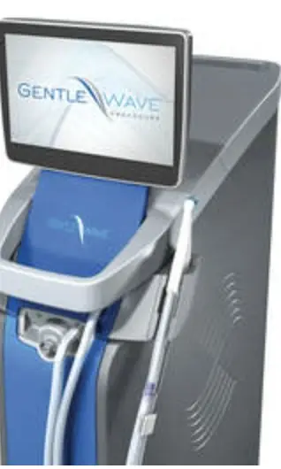
Cleaning the deepest, most complex parts of the root canal system takes real innovation. The GentleWave Procedure is an advanced alternative to standard root canal cleaning. It combines fluid dynamics with a broad range of sound waves to reach microscopic spaces and remove bacteria, debris and tissue.1
- Thorough cleaning of the root canal system, with less chance of failure over time.2
- Minimally invasive. A smaller opening preserves more of your natural tooth and keeps it stronger.3
- Often one visit. We can typically clean and fill the tooth in a single appointment.
Cone beam 3D imaging (CBCT)
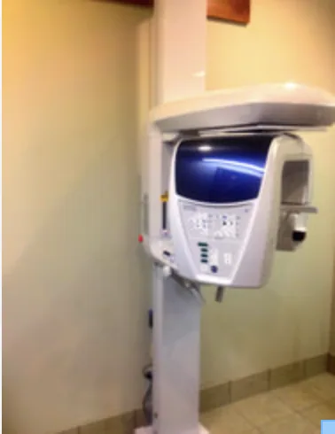
A cone beam CT takes a small three-dimensional image that can be sliced and rotated to diagnose problems and plan treatment. It uses very little radiation, about the same as three regular dental x-rays (roughly 15 µSv).
Before MRI and CT, doctors sometimes had to operate just to find a problem. 3D imaging does the same for teeth: we can often see the cause without opening the tooth. Cottonwood Endodontics was the first endodontic practice in Utah, and one of the first in the country, to use this technology, and Dr. Warren has lectured nationally and internationally on 3D imaging in endodontics.
Surgical operating microscope
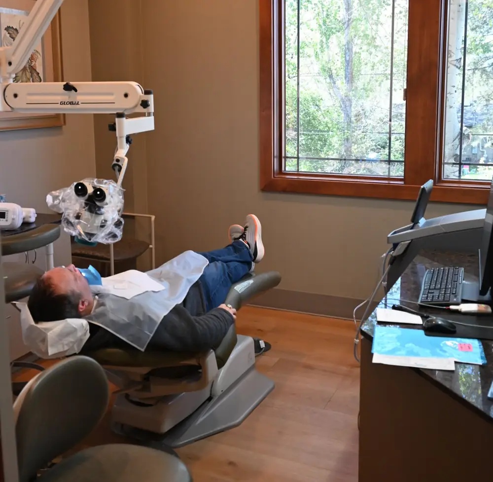
Root canal anatomy is often too small to see with the naked eye. We use a surgical operating microscope in every case. The magnification and light help us find and clean every canal, and in surgery they let us keep the surgical site small, which reduces discomfort and speeds healing.
Digital x-rays
All of our x-rays are digital. A small sensor captures the image using about one-tenth the radiation of film. Images appear on screen right away and can be enhanced for a clearer diagnosis.
1 Molina B et al. (2015) J Endod 41:1701–5. 2 Vandrangi P et al. (2015) Oral Health 72–86. 3 Sigurdsson A et al. (2016) J Endod 42:1040–48. GENTLEWAVE and the GENTLEWAVE logo are registered trademarks of Sonendo, Inc. and used under license.